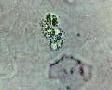
Leukocytes
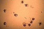
Leukocytes or white blood cells designate all the hemoglobin free blood cells. These cells belong to the reticuloendothelial system. Based on their nuclear aspect, white blood cells can be divided into two categories: mononuclear cells and polynuclear cells. Lymphocytes and monocytes are the principal mononuclear cells and polynuclear are subdivided into neutrophilic, eosinophilic and basophilic cells. In the urinary sediment, the term leukocyte is usually interpreted as polynuclear, mostly neutrophils. The reason for this situation is that the neutrophils are, by far, the most abundant leukocytes in urine. In a normal specimen, up to 6 or 7 neutrophils / high-power field can be observed. High neutrophil counts are usually related to an inflammation process.
Mononuclear leukocytes are occasionally seen. High mononuclear counts are usually related to a high blood count pathology, infiltrating the urinary space.
Under bright field microscopy and without staining, identification of the different types of white blood cells is almost impossible. The term leukocytes is indicated for the routine microscopy. The subclasses of leukocytes can be identified by staining with the Wright or PAP stain. Efficiency of these staining procedures is highly dependent of the conservation state of the cells. Staining with these represents a workload, not justified for the routine specimen.
Neutrophils
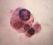
A polynuclear neutrophil (neutro) is a plurilobular nucleus bearing cell with a slightly granulated cytoplasm. Neutros have two types of granulations (lysozome) named azurophilic and specific. Neutros lysozomes havediverse enzyme activities, some being specific like the peroxydase. Neutrophils also possess a heterogenic group of hydrolase gathered under the term esterase. One of these esterases, the Naphtyl AS-D Chloroacetate esterase, seems to be specific for the polynuclear lineage. This activity is shared with the mastocytes and some macrophages (some think that this activity is due to phagocyted neutros). Since mastocytes are not seen in urine, and that the macrophage size and aspect is quite different from leukocytes, this activity is used in a specific staining procedure for leukocytes.
Because of a pycnotic nucleus or of an unfavorable refractive index of the urine, the plurilobulated nucleus are not always obvious. Acidification of the sediment, with one drop of 2% acetic acid enhances the contrast of the preparation.
 In the urinary sediment,
there are two types of neutrophils. The first type is the
usual named "Old" by Stamey. When
numerous, these cells are related to inflammation. In the urinary sediment,
there are two types of neutrophils. The first type is the
usual named "Old" by Stamey. When
numerous, these cells are related to inflammation. |
 The second type, named
"Fresh" by Stamey, and "Pale cells"
by Sternheimer, is bigger in size and resistant to some
stains. If the urine density is lower than 1,019 this
cell will demonstrate a brownian movement of its granules
wich will give a glittering cytoplasm. These cells are
then called glitter cells. For a time, these cells were
thought to be specific to pyelonephritis. Since these
were found in other conditions the accepted
interpretation is relating them to an active inflammation
process of the urinary tract. The second type, named
"Fresh" by Stamey, and "Pale cells"
by Sternheimer, is bigger in size and resistant to some
stains. If the urine density is lower than 1,019 this
cell will demonstrate a brownian movement of its granules
wich will give a glittering cytoplasm. These cells are
then called glitter cells. For a time, these cells were
thought to be specific to pyelonephritis. Since these
were found in other conditions the accepted
interpretation is relating them to an active inflammation
process of the urinary tract. |
Pus
 One
characteristic of the activated neutrophil is it's adherent
capacity. This characteristic is essential for the migration of
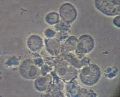
the cell. Because of this, neutrophils can easily aggregate. In
some cases, it is important not to confuse cell aggregation and
pus. Pus is formed of degenerated neutrophils (pyocytes) and
cellular debris compacted into a mass where cell identity is
lost. This discrimination is not commonly used with the urinary
sediment, so that many aggregates are reported as pus. The term
pus should be restrained to real pus.
One
characteristic of the activated neutrophil is it's adherent
capacity. This characteristic is essential for the migration of
the cell. Because of this, neutrophils can easily aggregate. In
some cases, it is important not to confuse cell aggregation and
pus. Pus is formed of degenerated neutrophils (pyocytes) and
cellular debris compacted into a mass where cell identity is
lost. This discrimination is not commonly used with the urinary
sediment, so that many aggregates are reported as pus. The term
pus should be restrained to real pus.
Eosinophils
This cell is different from the other polynuclear cells because of the affinity of its granules for the acid stains like eosin. To be able to distinguish this cell, it is absolutely necessary to stain. In a comparative study, by Nolan and Kelleher, the former has shown that the Hansel stain was superior to the Wright stain for eosinophiluria. The presence of urinary eosinophils is a useful indicator of acute allergic interstitial nephritis. Eosinophiluria is also seen in conditions other than nephritis.
Causes of eosinophiluria
| Acute allergic interstitial nephritis |
| Rapidly progressive glomerulonephritis |
| Acute glomerulonephritis |
| IgA nephropathy, Henoch-Schönlein purpura |
| Prostatitis |
| Schistosomiasis |
| Chronic pyelonephritis |
| Graft rejection (acute phase) |
Lymphocytes
Lymphocytes can occasionally be seen in a normal sediment. High counts have been reported in cases of acute allergic interstitial nephritis, rapidly evolutive glomerulonephritis, and graft rejection.
With bright field microscopy, the cell cannot be differentiated from the usual leukocytes. With the PAP stain, the cell is small, as a round nucleus with little cytoplasm.
Monocytes
In a study using monoclonal antibodies, a high number of urinary monocytes has been reported in cases of acute allergic interstitial nephritis and of rapidly evolutive glomerulonephritis. In necrosis, these were rare or absent.
Like the lymphocytes, monocytes cannot be identified with bright field microscopy. With the PAP stain, the cell is larger than a neutrophil and has a characteristic bean shaped nucleus.
Macrophages
 The macrophage is, after the fibroblast, the most
abundant cell in connective tissue. The activated macrophage is
difficult to describe because it has a very variable aspect. This
cell often presents itself with so many cytoplasmic inclusions
that the cell's structures are completely masked. Inclusions are
of several types, but the droplet is the most frequent. A classic
easy to identify macrophage, is the giant cell that contains one
or two smaller phagocyted cells in its cytoplasm. But this form
is exceptional; the majority of the macrophages are of average
size with a lot of inclusions.
The macrophage is, after the fibroblast, the most
abundant cell in connective tissue. The activated macrophage is
difficult to describe because it has a very variable aspect. This
cell often presents itself with so many cytoplasmic inclusions
that the cell's structures are completely masked. Inclusions are
of several types, but the droplet is the most frequent. A classic
easy to identify macrophage, is the giant cell that contains one
or two smaller phagocyted cells in its cytoplasm. But this form
is exceptional; the majority of the macrophages are of average
size with a lot of inclusions.
The macrophages are frequent in acute inflammations. The macrophage loaded with fat droplets is frequently found in many body fluids. These are usually related to a chronic inflammation process. In urine, when these droplets form a maltese cross when viewed between crossed polarized filters, the macrophages are then called oval fat bodies.