|
Type II granular casts
 Granular casts (type II)
are described by Lindner as hyaline matrix casts filled with
granules similar to cytoplasmic degeneration granules. There is a
relation between the structure of these granules and the granular
cytoplasm of the degenerating tubular cells. The causes of this
degeneration are unknown, but a proteinuria is a usual finding. A
protein overload could be responsible for the granular
degeneration of the tubular cells. The cytoplasm granulation
could then be integrated in a cast as free granules, as
cytoplasmic fragments, or as complete cells.
Granular casts (type II)
are described by Lindner as hyaline matrix casts filled with
granules similar to cytoplasmic degeneration granules. There is a
relation between the structure of these granules and the granular
cytoplasm of the degenerating tubular cells. The causes of this
degeneration are unknown, but a proteinuria is a usual finding. A
protein overload could be responsible for the granular
degeneration of the tubular cells. The cytoplasm granulation
could then be integrated in a cast as free granules, as
cytoplasmic fragments, or as complete cells.
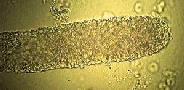
 Many of the
granular casts, when stained with PAP method, will show some
nucleus remnants and therefore, can be considered as a cellular
cast. These nuclear residues are not visible with unstained
specimens under bright field microscopy. In this condition, the
cast shows a homogenous granular texture typical of the type
II granular cast.
Many of the
granular casts, when stained with PAP method, will show some
nucleus remnants and therefore, can be considered as a cellular
cast. These nuclear residues are not visible with unstained
specimens under bright field microscopy. In this condition, the
cast shows a homogenous granular texture typical of the type
II granular cast.
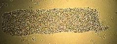
The texture varies from finely granular to coarsely granular. The granules size is often the same for all the casts on a specimen or on different specimens of the same patient, but is variable from one patient to another. Examples of this variation is shown in our picture collection. Coarse granules do not seem to be more clinically significant than the fine granules and are probably due to different synthesis conditions. These granular casts are non specific and represent a degradation of the tubular epithelia environments. A rare finding of these casts is considered physiological.
Type I granular casts
Lindner, Haber, and others have described the type I granular casts as a cast embedding cellular debris. This type I cast has a variable size granulation with a clumpy distribution. The leukocytes origin of the debris is suspected, but our attempt to stain these casts with the Naphtyl AS-D Chloro-acetate esterase gave deceiving results. We think that these casts are made of cellular debris of different kinds including degenerated leukocytes. The term "cellular debris cast" would be more appropriate and less confusing than the type I granular casts.
Muddy brown casts
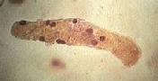
 These coarsely granular
casts are special by their dirty red-brown colorations (burnt
umber). The casts are rather large and heavily pigmented from
brown to almost black. The matrix is hyaline, with frequently one
end larger than the other.
These coarsely granular
casts are special by their dirty red-brown colorations (burnt
umber). The casts are rather large and heavily pigmented from
brown to almost black. The matrix is hyaline, with frequently one
end larger than the other.
The dirty brown color is due to pigments present within the granulation. These pigments seem to be oxydative degradation products of hemoglobin like methemoglobin and other products. The color is quite similar to air-exposed dried blood.
 The dirty brown color is not always easy
to distinguish from the orange red color of blood casts. The
drawing is an attempt to show the color difference between these.
The dirty brown color is not always easy
to distinguish from the orange red color of blood casts. The
drawing is an attempt to show the color difference between these.
The presence of large brown colored casts, alone, without any other anomaly, is suspicious. The context is very important in identification. Dirty brown casts are normally accompanied by hematuria, cellular casts, several tubular cells, often necrotic and pigmented and, if stasis is not recent, waxy casts. These casts must not be confused with plain hyaline casts rolled in amorphous urates.
The dirty brown casts are associated with acute tubular necrosis. This tubular necrosis is often of ischemic nature. These casts can also be seen in cases of massive intravascular hemolysis leading to renal failure.