In acid media non pH dependant In alkaline media |
Cystine
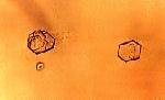 The cystine is seen as colorless
hexagonal plates. The solubility of the cystine is much larger in
alkaline urine, with the result that the former is rarely found
in alkaline urine. These colorless crystals can be difficult to
distinguish from the hexagonal plate form of uric acid crystals.
But, in this case, the examination of the crystals under
polarized light will probably show birefringent crystals with a
polarization color interference.
The cystine is seen as colorless
hexagonal plates. The solubility of the cystine is much larger in
alkaline urine, with the result that the former is rarely found
in alkaline urine. These colorless crystals can be difficult to
distinguish from the hexagonal plate form of uric acid crystals.
But, in this case, the examination of the crystals under
polarized light will probably show birefringent crystals with a
polarization color interference.
Cystine crystals is a clinically significant finding.
A confirmatory test is described elsewhere.
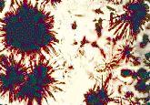
Leucine et tyrosine
Crystals of the amino acids leucine and tyrosine are very rarely seen in a urinary sediment. These crystals can be observed in some hereditary diseases like tyrosinosis and the "Maple syrup disease", but these conditions are very rare. The majority of cases where one finds these crystals are in patients with a serious hepatic problem, often in a terminal stage. In these cases, a concurrent presence of leucine and tyrosine is observed.
Leucine
|
Tyrosine
|
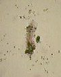
Bilirubin
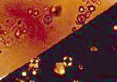
 Bilirubine crystallizes
in the urine as fine needles that regroup in a clump or as red
brown spheres. The clinical meaning is the same as the bilirubin
detected with the dipstick.
Bilirubine crystallizes
in the urine as fine needles that regroup in a clump or as red
brown spheres. The clinical meaning is the same as the bilirubin
detected with the dipstick.
Cholesterol
 Cholesterol
crystallizes as thin rectangular plates with one of the corners
(sometimes two or more) having a square notch. These crystals are
very slightly birefringent. The cause of the presence of
crystallized cholesterol is obscure. These crystals are seen in
degenerative kidney diseases and are thought to have an identical
clinical meaning as oval fat bodies. The presence of these
crystals is normally accompanied by a heavy proteinuria. These
crystals are very rare.
Cholesterol
crystallizes as thin rectangular plates with one of the corners
(sometimes two or more) having a square notch. These crystals are
very slightly birefringent. The cause of the presence of
crystallized cholesterol is obscure. These crystals are seen in
degenerative kidney diseases and are thought to have an identical
clinical meaning as oval fat bodies. The presence of these
crystals is normally accompanied by a heavy proteinuria. These
crystals are very rare.
Hemosiderin
 In a case of
intravascular hemolysis, a part of the free hemoglobin passes
through the glomerule. The former is then reabsorbed by the
tubular cells. The hemoglobin is then concentrated and
transformed slowly to a deep red brown pigmented granules, the
hemosiderin. These hemosiderin granules can be seen free, inside
tubular cells, and embedded in a cast. The free granules
agglomerate forming amorphous red brown deposit. A staining
procedure, based on the Roux reaction (Prussian blue), is
possible for the urinary sediment.
In a case of
intravascular hemolysis, a part of the free hemoglobin passes
through the glomerule. The former is then reabsorbed by the
tubular cells. The hemoglobin is then concentrated and
transformed slowly to a deep red brown pigmented granules, the
hemosiderin. These hemosiderin granules can be seen free, inside
tubular cells, and embedded in a cast. The free granules
agglomerate forming amorphous red brown deposit. A staining
procedure, based on the Roux reaction (Prussian blue), is
possible for the urinary sediment.
Ammonium biurates
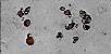
 The ammonium biurates, also called acid
ammonium urates, crystallize as a sphere with strias that reminds
a dried apple. Several crystals will show characteristic ox-horn
projections. The crystals are strongly birefringent. Ammonium
biurates are rarely seen in a fresh specimen. The former are
found in old specimens that turned alkaline.
The ammonium biurates, also called acid
ammonium urates, crystallize as a sphere with strias that reminds
a dried apple. Several crystals will show characteristic ox-horn
projections. The crystals are strongly birefringent. Ammonium
biurates are rarely seen in a fresh specimen. The former are
found in old specimens that turned alkaline.
Calcium carbonates
 The calcium carbonate
crystallizes as very small spheres. These spheres can be found
alone, in pair as dumbbell shape or in four units taking a cross
shape. These are strongly birefringent. Calcium carbonate
crystals are rare, probably because they are difficult to
distinguish from amorphous phosphates. Some authors report as
calcium carbonate what is recognized by others as amorphous
phosphates. The reason is that this crystal is found mixed with
amorphous phosphates thus forming a combined crystalluria of
homogeneous appearance. The clinical meaning of the calcium
carbonate is the same as amorphous phosphates.
The calcium carbonate
crystallizes as very small spheres. These spheres can be found
alone, in pair as dumbbell shape or in four units taking a cross
shape. These are strongly birefringent. Calcium carbonate
crystals are rare, probably because they are difficult to
distinguish from amorphous phosphates. Some authors report as
calcium carbonate what is recognized by others as amorphous
phosphates. The reason is that this crystal is found mixed with
amorphous phosphates thus forming a combined crystalluria of
homogeneous appearance. The clinical meaning of the calcium
carbonate is the same as amorphous phosphates.
Calcium sulfates
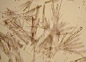 Calcium sulfate
crystallizes as thin plates with sharp ends. The plate can be
isolated or forming a rosette. These crystals are of little
clinical meaning.
Calcium sulfate
crystallizes as thin plates with sharp ends. The plate can be
isolated or forming a rosette. These crystals are of little
clinical meaning.