|
About transitional epithelium
From the pelvis to the beginning of the urethra, the walls of the urinary tract is lined with a stratified transitional epithelia. In the bladder, the epithelia is formed of around seven layers of cells. The deepest cells are cylindrical while the intermediate layer cells are variable. The surface cells are typically transitional. These cells are also named urothelial cells.
Transitional cells
 Transitional cells are
normal elements of the urinary sediment. The cell's shape changes
slightly according to the section of the lower urinary tract. The
typical bladder cells are round with a round centered nucleus. In
the cytological nomenclature, one speaks of: balloon cells,
umbrella cells, kite cells... All these designations correspond
to transitional cells originating from different levels: bladder,
uretere, pelvis.
Transitional cells are
normal elements of the urinary sediment. The cell's shape changes
slightly according to the section of the lower urinary tract. The
typical bladder cells are round with a round centered nucleus. In
the cytological nomenclature, one speaks of: balloon cells,
umbrella cells, kite cells... All these designations correspond
to transitional cells originating from different levels: bladder,
uretere, pelvis.
The presence of transitional cells is more frequent in the elderly population. Occasionally, because of morphological changes, the identification of transitional cells is difficult. Although these changes are not always associated to a pathology, atypical cells must be watched for in this prevalent population. Holmquist has demonstrated that a simple urinary routine sediment can play an important role in the early detection of TCC ( Transitionnal Cell Cancer).
Transitional epithelial fragments
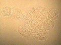 Contrarily
to the isolated cells, the presence of transitional epithelial
fragments is almost always associated to an abnormal situation.
In the majority of cases, the cells have a normal aspect and form
a thin sheet where it is easy to delimit each cell. These sheets
are said to have a brick-wall-like aspect. The presence of this
type of fragments can be the result of a urinary catheter or of
another condition that provokes an erosion of the surface of the
bladder's epithelium.
Contrarily
to the isolated cells, the presence of transitional epithelial
fragments is almost always associated to an abnormal situation.
In the majority of cases, the cells have a normal aspect and form
a thin sheet where it is easy to delimit each cell. These sheets
are said to have a brick-wall-like aspect. The presence of this
type of fragments can be the result of a urinary catheter or of
another condition that provokes an erosion of the surface of the
bladder's epithelium.
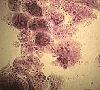 In some
sustained irritating conditions, transitional cells can become reactive. In these conditions, the cells and the nucleus increase
in size. The size of cells can be variable, but the ratio
nucleus/cytoplasm is well preserved. Binuclated cells can be seen.
In some
sustained irritating conditions, transitional cells can become reactive. In these conditions, the cells and the nucleus increase
in size. The size of cells can be variable, but the ratio
nucleus/cytoplasm is well preserved. Binuclated cells can be seen.
This situation is quite different from the atypical fragments as seen in a TCC of high grade. The finding of atypical urothelial fragments is an important element in the detection of an otherwise silent TCC.
| Characteristics of the atypical urothelial fragments (Holmquist) |
|---|
Cells
|
Nucleus
|
Nucleolus
|